
Visualisation of Single Cell Data and Segmentation of 3D Electron Microscopy Images
FAIR 1 Archive Notice
This page describes research activities and results from the FAIR 2019-2023 program period.
The content is retained for reference purposes and is no longer actively maintained. Information on this page, including research outputs, team members, contact details, collaborations and links, may be incomplete or outdated.
No direct successor use case has been defined within the FAIR 2024-2028 program. Please consult the current FAIR use cases for related research activities.
Use Case Description
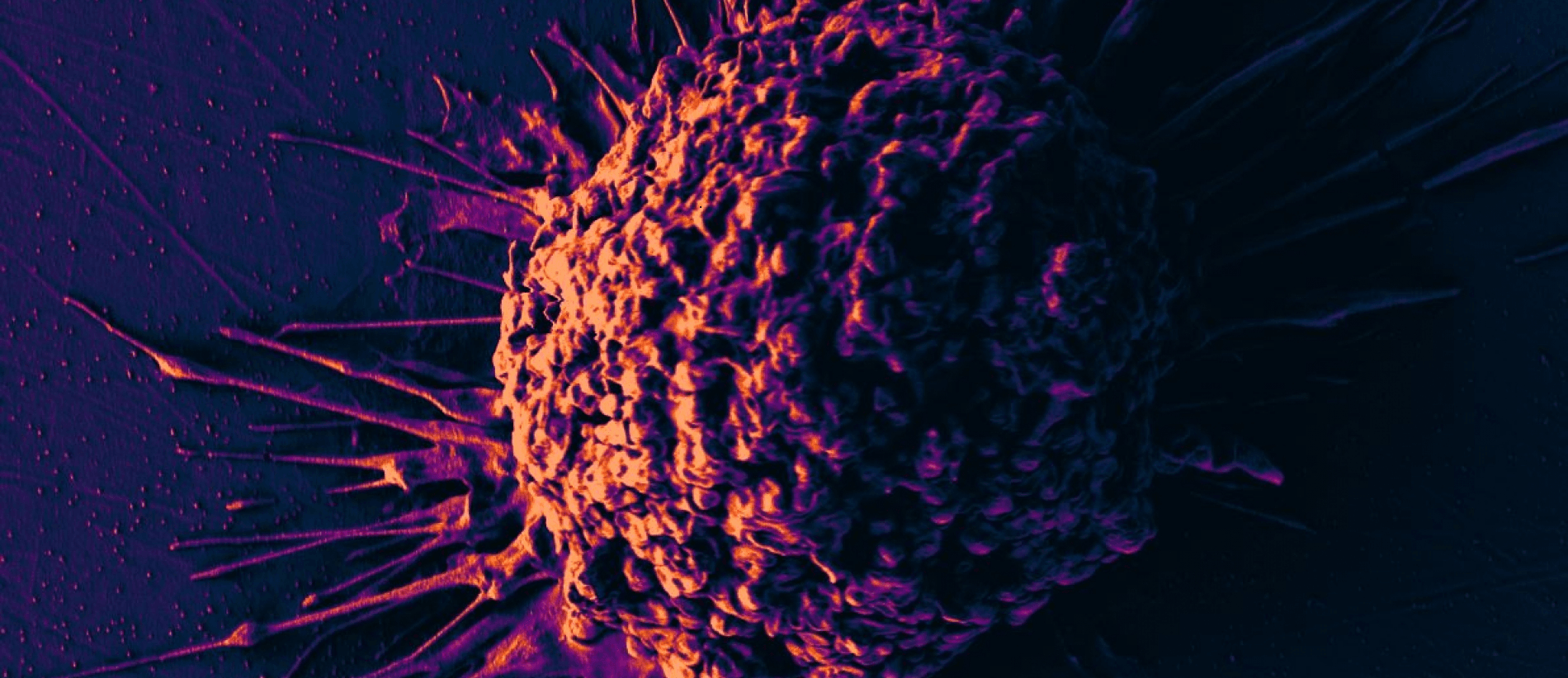
Cells are the basic units of life, and studying them holds immense significance for both medicine and the broader field of life sciences due to its ability to unravel the complexities of cellular behavior. Recent advances in high-throughput single-cell screening allows researchers to describe cells in unprecedented details, and this for massive amounts of cells. These novel technologies hold great promise to advance our understanding of cellular dynamics and functioning.
In medicine, understanding the intricacies of individual cells can revolutionize diagnostics and treatments, allowing for personalized medicine tailored to each patient's unique cellular profile. By analyzing single cells, researchers can identify subtle variations in gene expression, protein levels, and cellular interactions that underlie disease states, enabling the development of targeted therapies and more accurate prognoses.
In plant science and ecology, single-cell technologies play a pivotal role in elucidating the molecular mechanisms underlying plant growth, development, and responses to environmental stimuli. By dissecting the intricate cellular processes within plant tissues at the single-cell level, researchers can uncover the genetic and epigenetic factors influencing traits such as stress tolerance, yield, and nutrient uptake. This knowledge is invaluable for crop improvement efforts aimed at enhancing agricultural productivity and sustainability in the face of climate change and dwindling resources. Additionally, single-cell technologies offer insights into plant-microbe interactions, ecosystem dynamics, and biodiversity conservation.
Single-cell technologies offer insights into fundamental biological processes, shedding light on cellular heterogeneity, developmental trajectories, and tissue organization. Ultimately, delving into the intricacies of single cells not only enhances our understanding of human health and disease but also deepens our comprehension of the fundamental principles governing life itself.
The integration of artificial intelligence (AI) into single-cell technologies has revolutionized the fields of medicine, life sciences, plant science, and ecology, unlocking new insights and accelerating discoveries in cellular biology and ecology. AI algorithms excel at analyzing complex, high-dimensional datasets generated by single-cell technologies, enabling researchers to extract meaningful patterns, identify novel cell types, and unravel intricate cellular dynamics with unprecedented speed and accuracy. In medicine, AI-powered analysis of single-cell omics data facilitates the identification of disease biomarkers, prediction of treatment responses, and discovery of therapeutic targets, paving the way for personalized medicine and precision oncology. Overall, the synergy between AI and single-cell technologies empowers researchers to tackle longstanding challenges in biology and ecology, driving innovation and propelling scientific advancements in diverse fields.
Results-Artefacts
BioSegment: an automated, human-in-the-loop model for 3D-electron microscopy segmentation
Large 3D electron microscopy images require labor-intensive segmentation for further quantitative analysis. Recent deep learning segmentation methods automate this computer vision task, but require large amounts of labeled training data. We present BioSegment 2.0, a turnkey platform for experts to automatically process their imaging data and fine-tune segmentation models. It provides a user-friendly annotation experience, integration with familiar microscopy annotation software and a job queue for remote GPU acceleration. Various active learning sampling strategies are incorporated, with maximum entropy selection being the default. For mitochondrial segmentation, these strategies can improve segmentation quality by 10 to 15% in terms of intersection-overunion score compared to random sampling. Additionally, a segmentation of similar quality can be achieved using 25% of the total annotation budget required for random sampling
Software available at https://github.com/vibbic/biosegment
Modelling cell-cell communication with NicheNet
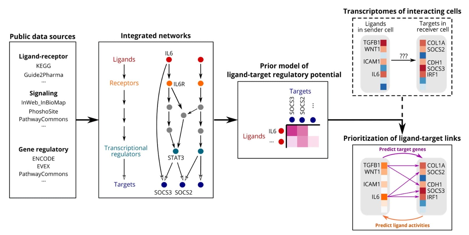
The goal of NicheNet is to study intercellular communication from a computational perspective. NicheNet uses human or mouse gene expression data of interacting cells as input and combines this with a prior model that integrates existing knowledge on ligand-to-target signaling paths. This allows to predict ligand-receptor interactions that might drive gene expression changes in cells of interest.
Press release: VIB scientists develop Twitter for cells.
Dyngen: a multi-modal simulation engine for single-cell omics
We developed dyngen, a novel, multi-modal simulation engine for studying dynamic cellular processes at single-cell resolution. dyngen is more flexible than current single-cell simulation engines, and allows better method development and benchmarking, thereby stimulating development and testing of novel computational methods.
Available at https://dyngen.dynverse.org/
Towards better flow cytometry data analysis in clinical practice with FlowSOM and PeacoQC
We developed several new algorithms for the automated analysis of single-cell cytometry data. This data is routinely used in clinical practice for immune monitoring. Using the FlowSOM ecosystem we offer state-of-the-art tools for automated population discovery, visualization and comparison. Our new PeacoQC tool advances the state-of-the-art in automated quality control for cytometry data.
Journal/Conference Publications
[1] Côrte-Real BF, Hamad I, Arroyo Hornero R, Geisberger S, Roels J, Van Zeebroeck L, Dyczko A, van Gisbergen MW, Kurniawan H, Wagner A, Yosef N, Weiss SNY, Schmetterer KG, Schröder A, Krampert L, Haase S, Bartolomaeus H, Hellings N, Saeys Y, Dubois LJ, Brenner D, Kempa S, Hafler DA, Stegbauer J, Linker RA, Jantsch J, Müller DN, Kleinewietfeld M. Sodium perturbs mitochondrial respiration and induces dysfunctional Tregs. Cell Metab. 2023 Feb 7;35(2):299-315
[2] Roels J, De Craemer AS, Renson T, de Hooge M, Gevaert A, Van Den Berghe T, Jans L, Herregods N, Carron P, Van den Bosch F, Saeys Y, Elewaut D. Machine Learning Pipeline for Predicting Bone Marrow Edema Along the Sacroiliac Joints on Magnetic Resonance Imaging. Arthritis Rheumatol. 2023 Dec;75(12):2169-2177
[3] Sang-aram, C. Browaeys, R., Seurinck, R. Saeys, Y. 2023 Spotless: a reproducible pipeline for benchmarking cell type deconvolution in spatial transcriptomics eLife12:RP88431
[4] Sonrel A, Luetge A, Soneson C, Mallona I, Germain PL, Knyazev S, Gilis J, Gerber R, Seurinck R, Paul D, Sonder E, Crowell HL, Fanaswala I, Al-Ajami A, Heidari E, Schmeing S, Milosavljevic S, Saeys Y, Mangul S, Robinson MD. Meta-analysis of (single-cell method) benchmarks reveals the need for extensibility and interoperability. Genome Biol. 2023 May 17;24(1):119
[5] Rombaut,B., Roels, J., Saeys, Y. (2022) BioSegment: Active Learning segmentation for 3D electron microscopy imaging. IAL@PKDD/ECML 2022: 7-26
[6] Karras P, Bordeu I, Pozniak J, Nowosad A, Pazzi C, Van Raemdonck N, Landeloos E, Van Herck Y, Pedri D, Bervoets G, Makhzami S, Khoo JH, Pavie B, Lamote J, Marin-Bejar O, Dewaele M, Liang H, Zhang X, Hua Y, Wouters J, Browaeys R, Bergers G, Saeys Y, Bosisio F, van den Oord J, Lambrechts D, Rustgi AK, Bechter O, Blanpain C, Simons BD, Rambow F, Marine JC. Author Correction: A cellular hierarchy in melanoma uncouples growth and metastasis. Nature. 2022 Nov;611(7934)
[7] Couckuyt A, Seurinck R, Emmaneel A, Quintelier K, Novak D, Van Gassen S, Saeys Y. Challenges in translational machine learning. Hum Genet. 2022 Sep;141(9):1451-1466
[8] Guilliams M, Bonnardel J, Haest B, Vanderborght B, Wagner C, Remmerie A, Bujko A, Martens L, Thoné T, Browaeys R, De Ponti FF, Vanneste B, Zwicker C, Svedberg FR, Vanhalewyn T, Gonçalves A, Lippens S, Devriendt B, Cox E, Ferrero G, Wittamer V, Willaert A, Kaptein SJF, Neyts J, Dallmeier K, Geldhof P, Casaert S, Deplancke B, Ten Dijke P, Hoorens A, Vanlander A, Berrevoet F, Van Nieuwenhove Y, Saeys Y, Saelens W, Van Vlierberghe H, Devisscher L, Scott CL. Spatial proteogenomics reveals distinct and evolutionarily conserved hepatic macrophage niches. Cell. 2022 Jan 20;185(2):379-396
[9] Emmaneel A, Quintelier K, Sichien D, Rybakowska P, Marañón C, Alarcón-Riquelme ME, Van Isterdael G, Van Gassen S, Saeys Y. PeacoQC: Peak-based selection of high quality cytometry data. Cytometry A. 2022 Apr;101(4):325-338
[10] Quintelier K, Couckuyt A, Emmaneel A, Aerts J, Saeys Y, Van Gassen S. Analyzing high-dimensional cytometry data using FlowSOM. Nat Protoc. 2021 Aug;16(8):3775-3801
[11] Cannoodt R, Saelens W, Deconinck L, Saeys Y. Spearheading future omics analyses using dyngen, a multi-modal simulator of single cells. Nat Commun. 2021 Jun 24;12(1):3942
[12] Wendrich JR, Yang B, Vandamme N, Verstaen K, Smet W, Van de Velde C, Minne M, Wybouw B, Mor E, Arents HE, Nolf J, Van Duyse J, Van Isterdael G, Maere S, Saeys Y, De Rybel B. Vascular transcription factors guide plant epidermal responses to limiting phosphate conditions. Science. 2020 Nov 13;370(6518)
[13] Browaeys R, Saelens W, Saeys Y. NicheNet: modeling intercellular communication by linking ligands to target genes. Nat Methods. 2020 Feb;17(2):159-162
Toolboxes developed
BIOSEGMENT (automated segmentation of 3D-EM data)
FlowSOM (Automated analysis of cytometry data)
NicheNet (Modelling intercellular communication)
PeacoQC (Automated quality control and preprocessing of cytometry data)
PhD
Robrecht Cannoodt "Modelling single-cell dynamics with trajectories and gene regulatory networks"
Robin Vandaele "Topological data analysis for machine learning"
Helena Todorov "Structure learning to unravel mechanisms of the immune system"
Outreach
Stories/Blogs
Who responds to medical treatments and who doesn't ?
External events and workshops
Workshop Translating Artificial Intelligence to the clinic
Conference Applied Bioinformatics in the Life Sciences (ABLS 2022)
Conference Emerging Technologies in Single cell research (SingleCell20)
Technology transfer and valorization
This use case has led to one spin-off activity, now rebranded as the Single-cell core facility at VIB. This facility provides services in single-cell data generation and analysis for academia and industry. During the last three years, this spin-off has grown from 5 to 11 FTE's.
Basic Use Case Info
Period
2019-2023
Contact
Yvan Saeys Yvan.Saeys@ugent.be
Research Groups
DAMBI-UGENT (Yvan Saeys, Ruth Seurinck)
AIDA-UGENT (Tijl De Bie, Jefrey Lijffijt)
GAIM-UGENT (Aleksandra Pizurica)
KERMIT-UGENT (Willem Waegeman, Bernard De Baets)
ADREM-UANTWERPEN (Kris Laukens)
IMEC (Roel Wuyts)
STADIUS (Bart De Moor)
External experts and User Groups
External experts
Alexander Botzki
Saskia Lippens
Martin Guilliams
Bert De Rybel
User group members
Alexander Botzki (VIB, Bioinformatics core)
Saskia Lippens (VIB, Bioimaging core)
What's next?
New use case Single Cell Technologies, including multi-modal and spatial single-cell data.