
Single Cell
Use Case Description
Single-cell technologies have been revolutionizing the life sciences, pharmaceutical and diagnostics industries during the last decade by offering unprecedented levels of details at which cells can be measured at single cell resolution (Aldridge, 2020). By offering a genome-wide readout, researchers can now measure genome sequences and gene expression at unprecedented scale, and this for tens of thousands of cells simultaneously. This is critical for analyzing the complexity of biological systems as found in many fields in the life sciences, including cancer, neurology, inflammation and stem cell research.
Single-cell technologies have emerged as the “next-generation microscopes”, offering unprecedented approaches to the field of precision medicine, which aims to tailor the treatment to the individual characteristics of the patient. By offering new ways of identifying biomarkers, stratifying patient groups based on underlying mechanistic differences and predicting treatment response they thus have far-reaching implications for the broad field of health sciences. By providing a readout at the whole genome level, single-cell “omics” approaches result in very detailed, but high-dimensional data, that crucially depend on novel advances in data science to yield novel biological insights (Lahneman, 2020).
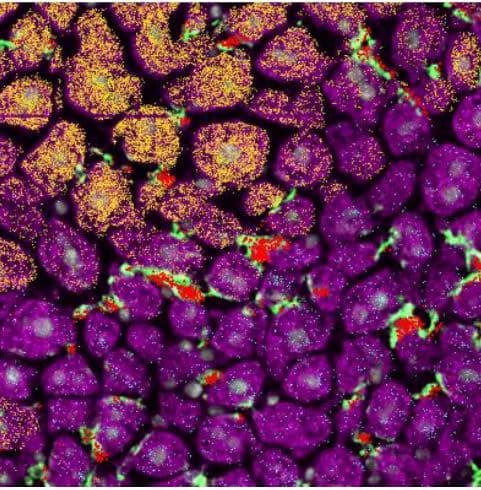
While the first wave of single-cell technologies mainly focused on scaling various “omics” readouts to single-cell resolution, current technologies take these approaches one step further. On the one hand, they allow multi-modal readouts (Macaulay, 2017) , combining several layers of omics information simultaneously at the single-cell level (e.g. transcriptomics, epigenetics, TCR/BCR sequencing, surface proteome,…), while on the other hand novel “spatial omics” technologies are a hybridization between omics readouts and imaging technology, providing us also with spatial information on these genomic readouts (Stahl, 2016). This allows scientists to map molecular readouts to very detailed spatial locations in a tissue, making it possible to better understand the structure and functioning of tissues, both in healthy and diseased states (Browaeys, 2020).
We define three AI-related challenges that will go beyond the current state-of-the-art, and will empower biologists with better tools to explore and interpret their data in a trustworthy fashion. These tools can then be used to gain new insights into how tissues function, providing the basis for developing more targeted treatments.
Multi-modal integration of single-cell omics data
Various omics and imaging technologies all shed a complementary light on cellular functioning, but current methods typically only focus on integrating a very limited number of types of omics data. We will explore novel integration methods that will allow combining "omics" and imaging data, and setup a framework to benchmark such integration methods, as such benchmarks are currently lacking.
Interactive data exploration and visualisation
To visualize current high-dimensional single-cell omics data, the current state-of-the-art still uses static visualizations based on dimensionality reduction methods such as tSNE and UMAP. We will go beyond this state-of-the-art and develop novel interactive exploration and visualization methods that can also take into account prior expert knowledge.
Automated functional annotation of cell types and cell states in space and time
Current single-cell atlas datasets provide a wealth of information to annotate cell types and states in an automated fashion for novel samples. We will design novel interpretable methods that can annotate various cellular aspects and provide insights into the gene regulatory and intercellular communication dynamics that underlie tissue functioning.
Results, Highlights & Research Outputs
Single-cell Open Problems: a living benchmark for single-cell analysis
Single-cell genomics has enabled the study of biological processes at an unprecedented scale and resolution. As single-cell technologies have become more prevalent, so has the development of new analysis tools, which has resulted in over 1,700 published algorithms (as of February 2024). Thus, there is an increasing need to continually evaluate which algorithm performs best in which context to inform best practices that evolve with the field.
We developed Open Problems, a living, extensive, community-guided platform including 12 current single-cell tasks that we envisage raising standards for the selection, evaluation and development of methods in single-cell analysis.
The Open Problems platform is an open-source, extensible, living benchmarking framework that enables quantitative evaluation of best practices in single-cell analysis. It combines a permissively licensed GitHub repository with community-defined tasks, an automated benchmarking workflow, and a website to explore the results. Currently, Open Problems includes 12 defined tasks, in which 81 datasets are used to evaluate 171 methods using 37 metrics. These tasks were defined by community engagement, including on the public GitHub repository, in weekly community meetings, and at a hackathon in March 2021 with over 50 participants. This broad involvement has already led to new benchmarking insights and best practice recommendations while improving and standardizing previously published benchmarks.
We envisage Open Problems’ community-defined standards for progress in single-cell data science raising the bar for the selection and evaluation of methods, providing targets for method innovation, and enabling developers without single-cell expertise to contribute to the field.
Software / Tool
Software available at https://openproblems.bio
Harpy: A library for single-cell spatial proteomics data analysis
Recent automated bioimaging platforms generate complex and high-throughput spatial omics data. Especially whole-slide images with dozens to hundreds of fluorescent protein image channels can be difficult to process and analyze at the single-cell level.
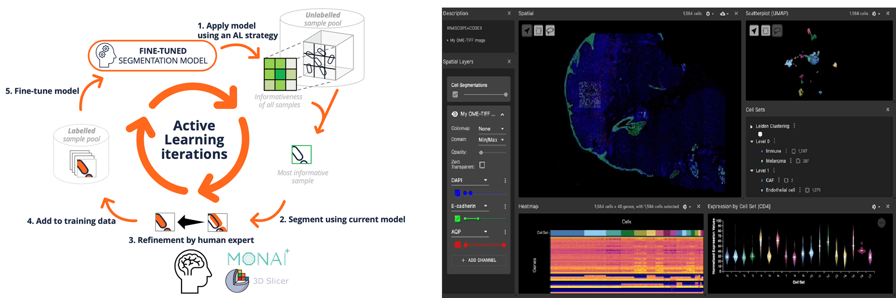
We present an analysis workflow with three key components: (1) secure dataset management using remote Big Data object storage and SpatialData, (2) scalable cell segmentation and feature calculation utilizing cellpose, Dask and high-performance computing, and (3) interactive expert-in-the-loop hyperparameter tuning and cell annotation using FlowSOM, napari and Jupyter notebooks. Feedback on data quality is provided through various plots, offering insights at each stage of the workflow such as overview of channel quality and comparisons of different segmentation methods.
Overall, this powerful yet user-friendly workflow empowers users to efficiently analyze large volumes of imaging data, significantly reducing the time and effort required to go from microscope to meaningful quantitative insights.
Software / Tool
Software available at https://github.com/saeyslab/harpy
Uncertainty-aware single-cell annotation with a hierarchical reject option
Automatic cell type annotation methods assign cell type labels to new datasets by extracting relationships from a reference RNA-seq dataset. However, due to the limited resolution of gene expression features, there is always uncertainty present in the label assignment. To enhance the reliability and robustness of annotation, most machine learning methods address this uncertainty by providing a full reject option, i.e. when the predicted confidence score of a cell type label falls below a user-defined threshold, no label is assigned and no prediction is made. As a better alternative, some methods deploy hierarchical models and consider a so-called partial rejection by returning internal nodes of the hierarchy as label assignment. However, because a detailed experimental analysis of various rejection approaches is missing in the literature, there is currently no consensus on best practices.
We evaluate three annotation approaches (i) full rejection, (ii) partial rejection, and (iii) no rejection for both flat and hierarchical probabilistic classifiers. Our findings indicate that hierarchical classifiers are superior when rejection is applied, with partial rejection being the preferred rejection approach, as it preserves a significant amount of label information. For optimal rejection implementation, the rejection threshold should be determined through careful examination of a method’s rejection behavior. Without rejection, flat and hierarchical annotation perform equally well, as long as the cell type hierarchy accurately captures transcriptomic relationships.
Software / Tool
Software available at https://github.com/Latheuni/Hierarchical_reject
Evaluation of out-of-distribution detection methods for data shifts in single-cell transcriptomics
Automatic cell-type annotation methods assign cell-type labels to new, unlabeled datasets by leveraging relationships from a reference RNA-seq atlas. However, new datasets may include labels absent from the reference dataset or exhibit feature distributions that diverge from it. These scenarios can significantly affect the reliability of cell type predictions, a factor often overlooked in current automatic annotation methods. The field of out-of-distribution detection (OOD), primarily focused on computer vision, addresses the identification of instances that differ from the training distribution. Therefore, the implementation of OOD methods in the context of novel cell type annotation and data shift detection for single-cell transcriptomics may enhance annotation accuracy and trustworthiness.
We evaluate six OOD detection methods: LogitNorm, MC dropout, Deep Ensembles, Energy-based OOD, Deep NN, and Posterior networks, for their annotation and OOD detection performance in both synthetical and real-life application settings. We show that OOD detection methods can accurately identify novel cell types and demonstrate potential to detect significant data shifts in non-integrated datasets. Moreover, we find that integration of the OOD datasets does not interfere with OOD detection of novel cell types.
Software / Tool
Software available at https://github.com/Latheuni/OOD_eval.git
Pattern or Artifact? Interactively Exploring Embedding Quality with TRACE
We introduce TRACE, a tool to analyze the quality of 2D embeddings of high-dimensional single-cell omics data generated through dimensionality reduction techniques. Dimensionality reduction methods often prioritize preserving either local neighborhoods or global distances, but insights from visual structures can be misleading if the objective has not been achieved uniformly. TRACE addresses this challenge by providing a scalable and extensible pipeline for computing both local and global quality measures. The interactive, browser-based interface allows users to explore various embeddings while visually assessing the pointwise embedding quality. The interface also facilitates in-depth analysis by highlighting high-dimensional nearest neighbors for any group of points and displaying high-dimensional distances between points. TRACE enables analysts to make informed decisions regarding the most suitable dimensionality reduction method for their specific use case, by showing the degree and location where structure is preserved in the reduced space.
Software / Tool
Software available at https://github.com/aida-ugent/TRACE
Interpretable scRNA-seq embeddings with multi-scale structure preservation using ViVAE and ViScore
Dimensionality reduction (DR) techniques are essential in single-cell transcriptomics: they capture important structures in expression data and produce low-dimensional representations for visualisation, discovery and downstream analysis.
Each DR method is designed to prioritise certain attributes of the original, high-dimensional data.
Drawing from recent developments in the fields of DR and deep learning, we introduce ViVAE: a structure-learning DR approach that achieves multi-scale preservation of structure and enhanced interpretability. Additionally, we advance DR evaluation methodology by introducing ViScore: a robust scoring and benchmarking framework. We address pitfalls of existing evaluation approaches and provide both unsupervised and supervised metrics which are scalable and fair.
Software / Tool
Software available at https://github.com/saeyslab/ViVAE
SpatialData: an open and universal data framework for spatial omics
Spatially resolved omics technologies are transforming our understanding of biological tissues. However, the handling of uni- and multimodal spatial omics datasets remains a challenge owing to large data volumes, heterogeneity of data types and the lack of flexible, spatially aware data structures. Here we introduce SpatialData, a framework that establishes a unified and extensible multiplatform file-format, lazy representation of larger-than-memory data, transformations and alignment to common coordinate systems. SpatialData facilitates spatial annotations and cross-modal aggregation and analysis, the utility of which is illustrated in the context of multiple vignettes, including integrative analysis on a multimodal Xenium and Visium breast cancer study.
Software / Tool
Software available at https://spatialdata.scverse.org/en/stable/
SCIP: A scalable, reproducible and open-source pipeline for morphological profiling of image cytometry and microscopy data
Imaging flow cytometry (IFC) provides single-cell imaging data at a high acquisition rate. It is increasingly used in image-based profiling experiments consisting of hundreds of thousands of multi-channel images of cells. Currently available software solutions for processing microscopy data can provide good results in downstream analysis, but are limited in efficiency and scalability, and often ill-adapted to IFC data.
In this work, we propose Scalable Cytometry Image Processing (SCIP), a Python software that efficiently processes images from IFC and standard microscopy datasets. We also propose a file format for efficiently storing IFC data. We showcase our contributions on two large-scale microscopy and one IFC datasets, all of which are publicly available. Our results show that SCIP can extract the same kind of information as other tools, in a much shorter time and in a more scalable manner.
Software / Tool
Software available at https://github.com/ScalableCytometryImageProcessing/SCIP
Generation of a T cell receptor, cytokine and cell repertoire synovial fluid atlas to define commonalities and dissimilarities between arthritic diseases through systems immunology approaches
Although different chronic arthritic diseases are defined by clinical factors like gender, psoriasis and auto-antibodies, the biology of inflamed joints while comparing the different diseases remains neglected. Here, after curating an inflamed joint derived T-cell receptor (TCR) database, our new TRIASSIC tool identified 66303 significantly convergent TCR clonotypes. Clustering TCR clonotypes showed that synovial fluid convergence clusters (SFCCs) characterized HLA-B27+ mediated diseases (spondyloarthritis, SpA, and enthesitis-related juvenile idiopathic arthritis, JIA-ERA), Lyme arthritis and oligoarticular JIA. Single-cell transcriptomics and bulk proteomics showed upregulated interferon type I and II and TNF-α pathways in oJIA. Collectively, our work showed that chronic arthritis is characterized by both disease-specific and broadly shared mechanisms.
Outputs & Resources
Publications
Marconato, L., Palla, G., Yamauchi, K.A. et al. SpatialData: an open and universal data framework for spatial omics. Nat Methods (2024)
Lauren Theunissen, Thomas Mortier, Yvan Saeys, Willem Waegeman, Uncertainty-aware single-cell annotation with a hierarchical reject option, Bioinformatics, Volume 40, Issue 3, March 2024
Edith Heiter, Liesbet Martens, Ruth Seurinck, Martin Guilliams, Tijl De Bie, Yvan Saeys, Jefrey Lijffijt: Pattern or Artifact? Interactively Exploring Embedding Quality with TRACE. ECML/PKDD (8) 2024: 379-382
Benjamin Rombaut, Lotte Pollaris, Chananchida Sang-aram, MV Cruysse, Robrecht Cannoodt, Frank Vernaillen, Arne Defauw, Julien Mortier, Mayar Ali, Kresimir Bestak, et al. Vib hackathon on spatial omics tools and methods. 2024. doi: https://doi.org/10.37044/osf.io/uf7d3.
D Novak, C de Bodt, P Lambert, JA Lee, S Van Gassen, Y Saeys. A framework for quantifiable local and global structure preservation in single-cell dimensionality reduction. bioRxiv, 2023.11. 23.568428
Chananchida Sang-aram, Robin Browaeys, Ruth Seurinck, Yvan Saeys (2024) Spotless, a reproducible pipeline for benchmarking cell type deconvolution in spatial transcriptomics eLife 12:RP88431.
Luecken MD, Gigante S, Burkhardt DB, Cannoodt R, Strobl DC, Markov NS, Zappia L, Palla G, Lewis W, Dimitrov D, Vinyard ME, Magruder DS, Andersson A, Dann E, Qin Q, Otto DJ, Klein M, Botvinnik OB, Deconinck L, Waldrant K; Open Problems Jamboree Members; Bloom JM, Pisco AO, Saez-Rodriguez J, Wulsin D, Pinello L, Saeys Y, Theis FJ, Krishnaswamy S. Defining and benchmarking open problems in single-cell analysis. Res Sq [Preprint]. 2024 Apr 4:rs.3.rs-4181617. doi: 10.21203/rs.3.rs-4181617/v1.
Artuur Couckuyt, Benjamin Rombaut, Yvan Saeys, Sofie Van Gassen, Efficient cytometry analysis with FlowSOM in Python boosts interoperability with other single-cell tools, Bioinformatics, Volume 40, Issue 4, April 2024, btae179
Pozniak J, Pedri D, Landeloos E, Van Herck Y, Antoranz A, Vanwynsberghe L, Nowosad A, Roda N, Makhzami S, Bervoets G, Maciel LF, Pulido-Vicuña CA, Pollaris L, Seurinck R, Zhao F, Flem-Karlsen K, Damsky W, Chen L, Karagianni D, Cinque S, Kint S, Vandereyken K, Rombaut B, Voet T, Vernaillen F, Annaert W, Lambrechts D, Boecxstaens V, Saeys Y, van den Oord J, Bosisio F, Karras P, Shain AH, Bosenberg M, Leucci E, Paschen A, Rambow F, Bechter O, Marine JC. A TCF4-dependent gene regulatory network confers resistance to immunotherapy in melanoma. Cell. 2024 Jan 4;187(1):166-183.e25.
Hemberg, Martin; Marini, Federico; Ghazanfar, Shila; Al Ajami, Ahmad; Abassi, Najla; Anchang, Benedict; Benayoun, Bérénice A.; Cao, Yue; Chen, Ken; Cuesta-Astroz, Yesid; DeBruine, Zach; Dendrou, Calliope A.; De Vlaminck, Iwijn; Imkeller, Katharina; Korsunsky, Ilya; Lederer, Alex R.; Meysman, Pieter; Miller, Clint; Mullan, Kerry; Ohler, Uwe; Patikas, Nikolaos; Schuck, Jonas; Siu, Jacqueline H. Y.; Triche Jr, Timothy J.; Tsankov, Alex; van der Laan, Sander W.; Yajima, Masanao; Yang, Jean; Zanini, Fabio; Jelic, Ivana. “Insights, opportunities and challenges provided by large cell atlases.” arXiv preprint, arXiv:2408.06563 (2024).
Postovskaya A, Vercauteren K, Meysman P, Laukens K. tcrBLOSUM: an amino acid substitution matrix for sensitive alignment of distant epitope-specific TCRs. Brief Bioinform. 2024 Nov 22;26(1):bbae602
My K. Ha, Vincent Van Deuren, Carlos Alberto de Carvalho Fraga, An Hotterbeekx, Joke Dehoorne, Thomas Renson, Elke Geens, Nicolaas Aerts, Christiaan Heusdens, Maria Kuznetsova, Margot Van Mechelen, Michaël Vanden Bulcke, Ilse Hoffman, RuthWittoek, Eva De Wachter, Aurelie Andrzejewski, Esther Bartholomeus, Nick de Vrij, Arvid Suls, Eva Lion, Stijn Vanhee, Wim Adriaensen, Kerry Mullan, Helder Nakaya, Samir Kumar-Singh, Kris Laukens, Rik Joos, Pieter Meysman, Benson Ogunjimi. Generation of a T cell receptor, cytokine and cell repertoire synovial fluid atlas to define commonalities and dissimilarities between arthritic diseases through systems immunology approaches. bioRxiv 2025.01.10.632345; doi: https://doi.org/10.1101/2025.01.10.632345
Theunissen L, Mortier T, Saeys Y, Waegeman W. Evaluation of out-of-distribution detection methods for data shifts in single-cell transcriptomics. Brief Bioinform. 2025 May 1;26(3):bbaf239
Luecken MD, Gigante S, Burkhardt DB, Cannoodt R, Strobl DC, Markov NS, Zappia L, Palla G, Lewis W, Dimitrov D, Vinyard ME, Magruder DS, Andersson A, Dann E, Qin Q, Otto DJ, Klein M, Botvinnik OB, Deconinck L, Waldrant K; Open Problems Jamboree Members; Bloom JM, Pisco AO, Saez-Rodriguez J, Wulsin D, Pinello L, Saeys Y, Theis FJ, Krishnaswamy S. Defining and benchmarking open problems in single-cell analysis Nat Biotechnol 43, 1035–1040 (2025)
Lippeveld M, Peralta D, Filby A, Saeys Y. SCIP: A scalable, reproducible and open-source pipeline for morphological profiling of image cytometry and microscopy data. Cytometry A. 2024 Nov;105(11):816-828
Lippeveld,M., Peralta, D., Vardi, A. Vincent, F., Saeys, Y. (2025) Morphological profiling of imaging flow cytometry data uncovers heterogeneity in infected Gephyrocapsa huxleyi cultures. Cytometry A. 2025 Jul;107(7):438-449
Software & Tools
Single-cell Open Problems (A living and extensible benchmark for single-cell omics)
HARPY (single-cell Spatial Proteomics data analysis)
Hierarchical_reject(uncertainty-aware cell type annotation with reject option)
TRACE(interactive exploration of embedding quality)
ViVAE(multi-scale preservation structure learning)
ViScore(benchmarking framework for dimensionality reduction)
SpatialData(open and universal data framework for spatial omics)
OOD_eval (Out of distribution detection for single-cell transcriptomics)
SCIP (Scalable processing of imaging cytometry data)
PhD Dissertations
Benjamin Rombaut "An expert-guided pipeline for the analysis of spatial proteomics"
Yentl Van den Berghe "Image processing and quantitative image analysis of correlative light and electron microscopy data"
Chananchida Sang-aram "Modelling intercellular communication in time and space"
David Novak "Incorporating deep learning concepts in interpretable analyses of single-cell data"
Maxim Lippeveld "Development of scalable machine learning pipelines for image cytometry data"
Louise Deconinck "Alignment of cell developmental trajectories"
Edit Heiter "Interactive embeddings of high-dimensional data"
Laura Hajzoková "Interpretable and explorable single-cell embeddings"
Karen Herreman "Machine learning for spatial multi-omics data integration"
Xander Wieme "Interpretable machine learning models for graph-based neural networks"
Anna Postovskaya "T-cell receptor - epitope recognition models"
Sebastiaan Valkiers "Decoding the T cell receptor landscape in multiple sclerosis through sequence clustering and neighbor enrichment analysis"
Fuyin Lai "Pattern mining for understanding learned representations"
Outreach, Media & Events
Stories / Blogs
External Events & Workshops
Team & Contact
Contact
Use Case Lead(s)
Yvan Saeys, UGent / DAMBI
Principal Investigators / AI Expert Leads
Yvan Saeys, UGent / DAMBI
Research Groups
DAMBI-UGENT (Yvan Saeys, Ruth Seurinck)
AIDA-UGENT (Tijl De Bie, Jefrey Lijffijt)
GAIM-UGENT (Aleksandra Pizurica)
KERMIT-UGENT (Willem Waegeman)
ADREM-UANTWERPEN (Kris Laukens)