
Medical Imaging
Use Case Description
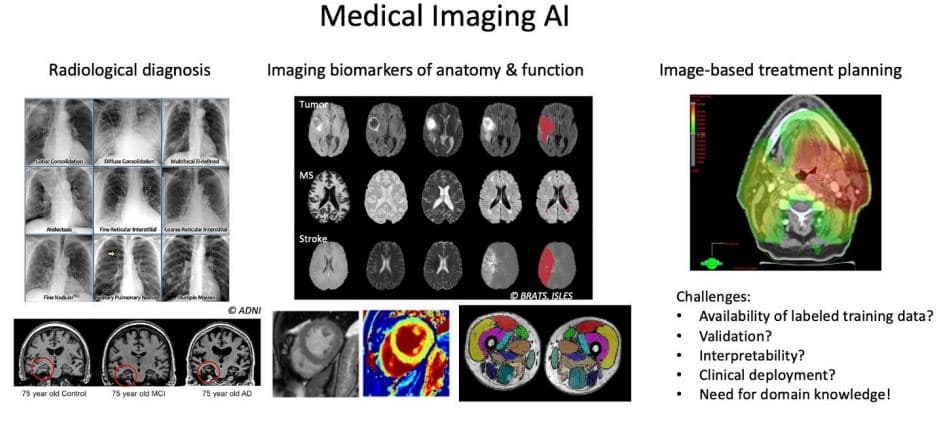
Medical imaging plays an essential role in many disciplines of medicine (Oncology, Cardiology, Neurology…) for diagnosis, therapy planning and follow-up, and in biomedical research. Various modalities based on different physical principles (RX, CT, MRI, US, PET…) are available in clinical practice that provide complementary information. Visual interpretation of the images by a trained medical expert is necessary subjective and many clinical applications would benefit from a more objective, quantitative analysis of the relevant image information. However, while manual analysis of medical images is too tedious to be practical in clinical routine, automated medical image analysis is complicated by the complexity of the images (3D, ambiguity due to limited resolution, lack of contrast and artifacts, variation in imaging protocols and image quality), of the imaged scenes (anatomical objects with complex 3D structure subject to normal variability and pathological abnormalities) and of the applications themselves, which are very diverse, multimodal, multitemporal, with clinical requirements evolving with continuous advances in medical imaging technology.
Computational strategies for medical image analysis using conventional analytical model-based strategies are limited by the underlying model assumptions made about the objects of interest (photometry, shape, context). Purely data driven methods on the other hand, in particular AI models based on deep learning, have shown great promise in various medical imaging tasks (detection, segmentation, quantification, fusion, but also image reconstruction and enhancement). However, the real-world performance of medical imaging AI models based on deep learning critically depends on the amount and relevance of the training data used, which is necessarily restricted in medical imaging applications due to the significant manual expert effort required to generate properly annotated ground truth data and which is also subject to intra- and inter-observer variability. Moreover, clinical adoption of medical imaging AI models, which often operate as a black box, requires that they are explainable and interpretable if their results will be used to support clinical decisions for diagnosis and treatment with direct impact on the patient.
Finally, while there is a strong clinical interest in the potential benefits of well-validated medical imaging AI models in various medical disciplines, their true clinical benefit can only be evaluated when they are effectively implemented and integrated in the (existing or adapted) clinical workflow in routine practice. Such integration is complicated not so much by technological difficulties (interfacing with the existing hospital information systems such as EPR and PACS and with cloud computing facilities) as by regulatory hurdles (in particular the medical device regulations - MDR).
In this use case, we aim at overcoming these challenges beyond the current state-of-the-art in order to enable unleashing the full potential of medical imaging AI tools in clinical practice in various applications. To this end, generic methodological advances in AI, as investigated in the generic research challenges in this project, are applied and adopted to medical imaging data in specific applications and augmented with extensive domain knowledge in close collaboration with clinical experts for learning reliable, interpretable image-based models from limited training data. The application-driven research within this use case is largely performed within the Medical Imaging Research Center of KU Leuven/UZ Leuven, which facilitates the access to state-of-the-art imaging data and expertise.
Objectives
To investigate the clinical benefit of AI-tools in the clinical setting, different medical imaging applications will be considered in this use case, involving mainly CT and MRI images, and primarily related to the quantification of imaging biomarkers of disease (e.g. measurement of size, volume, function, tissue parameters, lesion load…) and the use of these images for treatment planning, in particular future adaptive radiotherapy.
AI-tools for radiotherapy planning
This involves the development, initial evaluation and validation of clinical benefit of contouring and dose mapping methods for automating treatment planning in radiotherapy (RT) to enable fast replanning and online treatment adaptation, using the conventional planning CT, additional PET and MRI images if available and daily CBCT images as input. Initial models based on planning CT are already (in part) available and will be extended to be applicable to daily CBCT as well. Daily imaging with CBCT provides information on (significant) anatomical changes that may require replanning of the treatment. Final aim is the integration of different AI-tools in an end-to-end pipeline for automated fast replanning, enabling online treatment adaptation in the routine clinical workflow.
AI-tools for quantification of anatomy & function
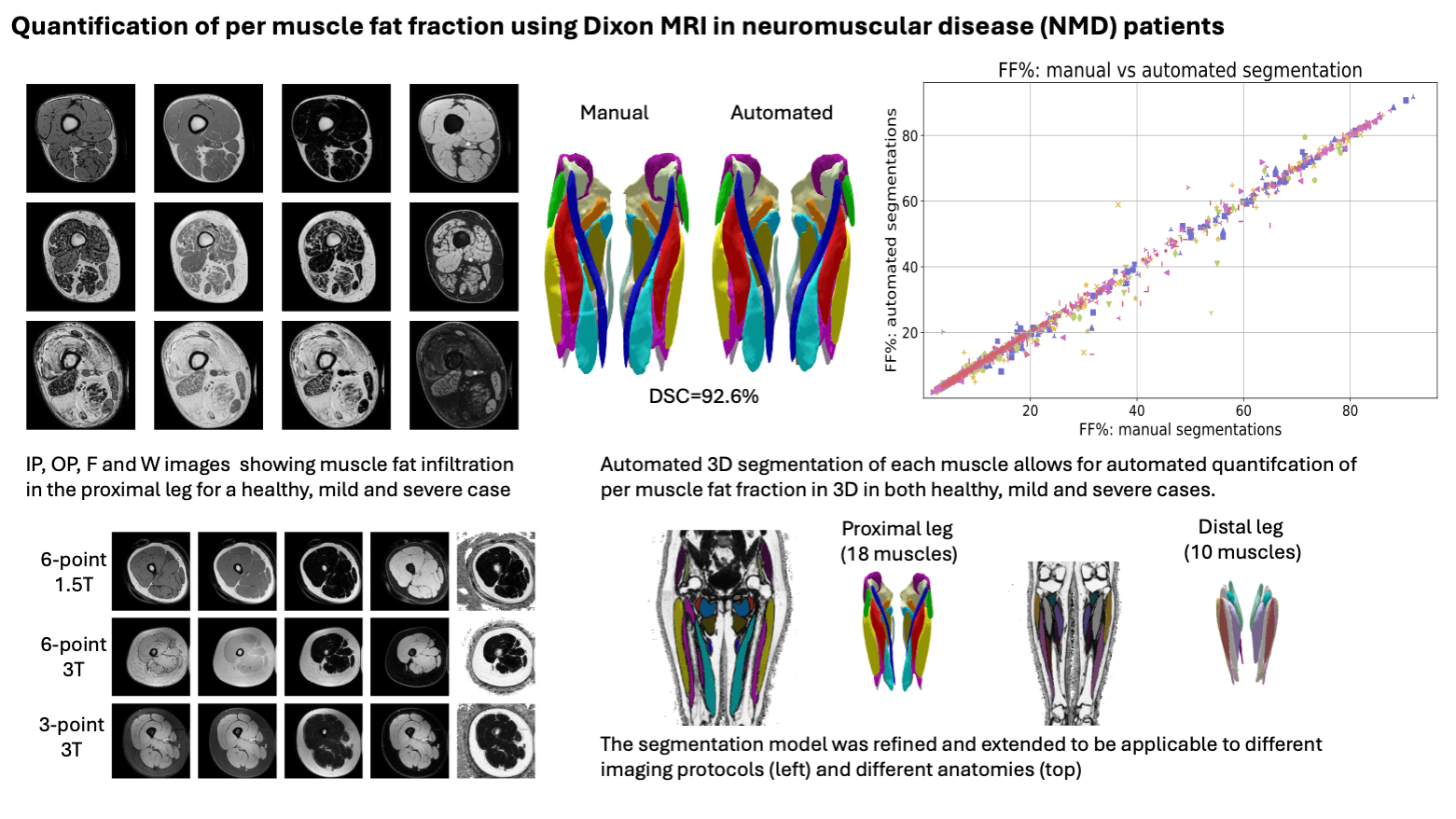
Targeted applications will evolve during the course of the project depending on clinical interest. Currently targeted applications involving MRI include the quantification of cardiac function from cardiac MRI, muscle fat fraction quantification in different body parts to evaluate disease progress in muscular dystrophy patients, microstructure analysis of the brain using DW-MRI, and the detection of lesions in whole-body DW-MRI. Applications involving CT include volumetry and anatomical modeling for surgery planning (including 3D printing). Different applications involve different domain knowledge but need to address similar challenges, such that progress in one application can benefit other applications as well, in particular: normalization of input data in view of variability in imaging protocols, fusion of images with complementary information (e.g. SA/LA views in cardiac MRI, multi-parametric MRI images), and incorporation of domain knowledge related to photometry, geometry, and context as flexible constraints.
Implementation, validation and deployment of AI-tools in the clinical setting
While deep learning is the current state-of-the-art in medical image analysis and has been shown to reach clinically relevant performance in several applications, the gap between the lab and the clinic is still large. Deployment of well-validated (in-house developed) AI tools in clinical practice is complicated by technological challenges related to the effective implementation and integration of these tools in the clinical workflow. We are closely collaborating with clinical partners and with the IT department of UZ Leuven to overcome these challenges. Generic solutions will be developed that are applicable to different applications operating along similar workflows using reusable components for patient image retrieval, anonymization, image preprocessing, interfacing with the AI-tool (running locally or in the cloud), post-processing of results and communication with other hospital IT components (PACS, EPR). Another major hurdle to overcome are the regulatory requirements imposed on software as a medical device (MDR), especially when these involve AI-based tools. We will investigate, together with the clinical stakeholders, for some specific clinical use cases the practical implications of these requirements for our research.
Results, Highlights & Research Outputs
MRI-based quantification of muscle fat fraction
An automated pipeline for MRI-based quantification of muscle fat fraction was developed in collaboration with Prof. Kristl Claeys, KU Leuven, Dept. Neurosciences, Laboratory for Muscle Diseases and Neuropathies, and UZ Leuven, Dept. Neurology.
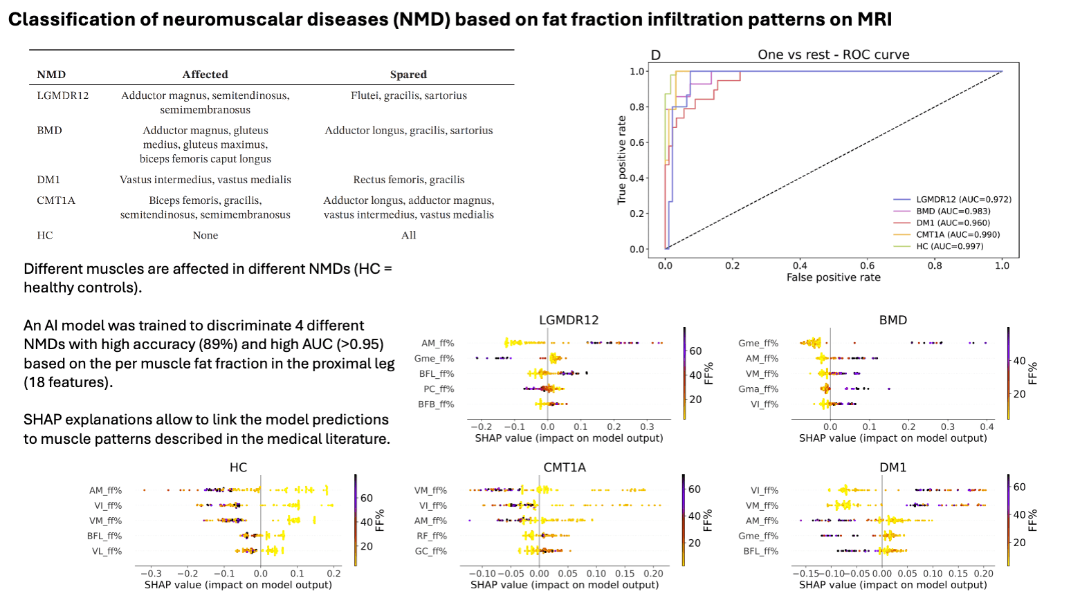
The quantified per muscle fat fractions allow to discriminate different types of neuromuscular diseases, affecting different muscles in the proximal leg, with high accuracy.
Outputs & Resources
Publications
Tilborghs, S., Liang, T., Raptis, S., Ishikita, A., Budts, W., Dresselaers, T., Bogaert, J., Maes, F., Wald, R.M., Van De Bruaene, A. (2024). Automated biventricular quantification in patients with repaired tetralogy of Fallot using a 3D deep learning segmentation model. Journal of Cardiovascular Magnetic Resonance. Sep 11:101092.
Rakic, M., Turco, F., Weng, G., Maes, F., Sima, D.M., Slotboom, J. (2024). Deep learning pipeline for quality filtering of MRSI spectra. NMR IN BIOMEDICINE, 37 (7). doi: 10.1002/nbm.5012
De Wel, B., Iterbeke, L., Huysmans, L., Peeters, R., Goosens, V., Dubuisson, N., van den Bergh, P., Van Parijs, V., Remiche, G., De Waele, L., Maes, F., Dupont, P., Claeys, K.G. (2024). Lessons for future clinical trials in adults with Becker muscular dystrophy: Disease progression detected by muscle magnetic resonance imaging, clinical and patient-reported outcome measures. EUROPEAN JOURNAL OF NEUROLOGY, 31 (7). doi: 10.1111/ene.16282
De Wel, B., Huysmans, L., Peeters, R., Ghysels, S., Byloos, K., Putzeys, G., Maes, F., Dupont, P., Claeys, K.G. (2024). Test-retest reliability and follow-up of muscle magnetic resonance elastography in adults with and without muscle diseases. JOURNAL OF CACHEXIA SARCOPENIA AND MUSCLE. doi: 10.1002/jcsm.13528
Sinonquel, P., Eelbode, T., Pech, O., De Wulf, D., Dewint, P., Neumann, H., Antonelli, G., Iacopini, F., Tate, D., Lemmers, A., Pilonis, N.D., Kaminski, M.F., Roelandt, P., Hassan, C., Ingrid, D., Maes, F., Bisschops, R. (2024). Clinical consequences of computer-aided colorectal polyp detection. GUT. doi: 10.1136/gutjnl-2024-331943
Koukoutegos, K., Maes, F., Bosmans, H. (2024). Cascade UNets for Kidney and Kidney Tumor Segmentation. In: N. Heller, A. Wood, F. Isensee, T. Radsch, R. Teipaul, N. Papanikolopoulos, C. Weight (Eds.), KIDNEY AND KIDNEY TUMOR SEGMENTATION, KITS 2023: vol. 14540, (107-113). Presented at the 3rd International Challenge on Kidney and Kidney Tumor Segmentation (KiTS), CANADA, Vancouver, 08 Oct 2023. ISBN: 978-3-031-54805-5. doi: 10.1007/978-3-031-54806-2_15